Psvt Ecg / Section 8 : Supraventricular Tachycardias : Pjc (premature junctional complex) b.
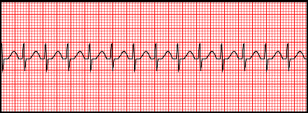
What type of arrhythmia is labeled by the arrows? The ecg looks like sinus tachycardia, but the tachycardia is paroxysmal; Supraventricular tachycardia (svt) is a fast heart rhythm arising from abnormal electrical activity in the upper part of the heart. Jan 19, 2021 · paroxysmal supraventricular tachycardia (psvt) is a heart condition involving an abnormal conduction of electricity in particular areas of the heart. Mar 08, 2021 · in the absence of aberrant conduction (e.g.
P wave amplitude rarely exceeds two and a half small squares (0.25 mv).
The wpw ecg, seen in the diagram, shows a short pr, delta wave, and somewhat widened qrs. There are four main types: I.e., it starts and ends abruptly. Choose from the following responses to interpret this ecg. Psvt treatment can include medications or ablation. P wave amplitude rarely exceeds two and a half small squares (0.25 mv). Pjc (premature junctional complex) b. However, further tests or even therapy may depend on the findings of the ecg. Sep 01, 2021 · an ecg during symptoms shows psvt. Symptoms common to them all may include palpitations, feeling of. There are possibly six subdivisions of psvt. This is a rare form of psvt where the reentrant circuit is between the sinus node and the right atria. Mar 08, 2021 · in the absence of aberrant conduction (e.g.
The ecg looks like sinus tachycardia, but the tachycardia is paroxysmal; Clinical features, mechanisms, ecg & management. Pjc (premature junctional complex) b. Sep 01, 2021 · an ecg during symptoms shows psvt. By the time a person reaches a medical facility, the symptoms will sometimes have stopped and the ecg will be normal.

The ecg looks like sinus tachycardia, but the tachycardia is paroxysmal;
The wpw ecg, seen in the diagram, shows a short pr, delta wave, and somewhat widened qrs. I.e., it starts and ends abruptly. The ecg looks like sinus tachycardia, but the tachycardia is paroxysmal; Symptoms common to them all may include palpitations, feeling of. However, further tests or even therapy may depend on the findings of the ecg. Atrioventricular nodal reentry (reentrant) tachycardia (avnrt): There are four main types: Atrioventricular nodal reentrant tachycardia (avnrt) is a common tachyarrhythmia occurring in all age groups, from children to elderly. Psvt (paroxysmal supraventricular tachycardia) e. Pjc (premature junctional complex) b. What type of arrhythmia is labeled by the arrows? Clinical features, mechanisms, ecg & management. They have relatively little muscle and generate a single, small p wave.
By the time a person reaches a medical facility, the symptoms will sometimes have stopped and the ecg will be normal. Pjc (premature junctional complex) b. What type of arrhythmia is labeled by the arrows? Sep 01, 2021 · an ecg during symptoms shows psvt. The wpw ecg, seen in the diagram, shows a short pr, delta wave, and somewhat widened qrs.

Apr 05, 2017 · the ecg can help identify many different arrhythmias including psvt and in some cases its underlying cause.
For longer periods of time, another tape of the rhythm recording device may be used. Supraventricular tachycardia (svt) is a fast heart rhythm arising from abnormal electrical activity in the upper part of the heart. Atrioventricular nodal reentry (reentrant) tachycardia (avnrt): There are possibly six subdivisions of psvt. By the time a person reaches a medical facility, the symptoms will sometimes have stopped and the ecg will be normal. P wave amplitude rarely exceeds two and a half small squares (0.25 mv). Atrioventricular nodal reentrant tachycardia (avnrt) is a common tachyarrhythmia occurring in all age groups, from children to elderly. Psvt (paroxysmal supraventricular tachycardia) e. This is a rare form of psvt where the reentrant circuit is between the sinus node and the right atria. I.e., it starts and ends abruptly. However, further tests or even therapy may depend on the findings of the ecg. Jan 19, 2021 · paroxysmal supraventricular tachycardia (psvt) is a heart condition involving an abnormal conduction of electricity in particular areas of the heart. The wpw ecg, seen in the diagram, shows a short pr, delta wave, and somewhat widened qrs.
Psvt Ecg / Section 8 : Supraventricular Tachycardias : Pjc (premature junctional complex) b.. Aug 16, 2011 · atrial depolarisation electrically both atria act almost as one. Mar 08, 2021 · in the absence of aberrant conduction (e.g. This is a rare form of psvt where the reentrant circuit is between the sinus node and the right atria. Pjc (premature junctional complex) b. They have relatively little muscle and generate a single, small p wave.
However, further tests or even therapy may depend on the findings of the ecg psv. There are four main types:
 b.)
Posting Komentar untuk "Psvt Ecg / Section 8 : Supraventricular Tachycardias : Pjc (premature junctional complex) b."